Ureteral Stricture

Restoring urine flow in the urinary tract

Ureteral stricture can be congenital or caused by trauma. Not addressing it, especially when symptoms show up, can be fatal. Early intervention will help prevent renal damage and reduce urinary symptoms.

The ureter transports urine from the kidney, where it collects in a funnel-shaped area called the renal pelvis. It then passes behind other organs in the abdomen and then down to the bladder. A stricture may develop at different points along the ureter. Where the ureter meets the kidney (ureteropelvic junction) and where it enters the bladder (ureteral orifice) are common locations where narrowing can occur.

When this tube becomes narrowed, urine cannot flow normally. Over time, this blockage can cause urine to back up into the kidney, leading to swelling (hydronephrosis), infection, and potential kidney damage if left untreated.

Diagnosis and Treatment Options

Diagnosing a ureteral stricture involves clinical evaluation and imaging tests. Doctors begin with a detailed medical history and physical exam, focusing on symptoms such as flank pain, blood in the urine, or recurrent infections. Past surgeries, radiation exposure, or prior kidney stones are also important clues.

Common Causes

Ureteral strictures can develop for several reasons. In some cases, the narrowing is present at birth (congenital), and in others, it develops later due to injury, inflammation, or other medical treatments.

  • Congenital narrowing – Some individuals are born with an abnormally narrow ureter. It can be identified in childhood, but sometimes it’s discovered later.
  • Surgical trauma – Procedures involving the urinary tract or nearby organs may lead to scar tissue formation that causes narrowing of the ureter.
  • Kidney stones – Passing a stone or undergoing procedures to remove one can irritate or damage the ureter.
  • Infections and inflammation – Recurrent urinary tract infections and other health conditions can cause scarring.
  • Radiation therapy – Treatment for cancers in the pelvis can damage ureteral tissue.
  • Tumors or external compression – Growths in or near the ureter can press on it and restrict urine flow.
  • Retroperitoneal fibrosis – A condition that causes scar tissue buildup in the back of the abdomen, potentially narrowing the ureter.

Imaging and diagnostic tests may include:

  • Ultrasound to detect kidney swelling (hydronephrosis)
  • CT urography provides detailed images of the urinary tract and helps identify the location and severity of the narrowing
  • MRI is used when radiation exposure should be limited or when more soft tissue detail is needed
  • Retrograde pyelography is an imaging test using contrast dye to directly visualize the ureter
  • Ureteroscopy uses a small camera to look inside the ureter
  • Urine tests to check for infection or other abnormalities

Imaging and testing help identify the cause, location, and severity of the stricture, and treatment options depend on those as well as the patient’s symptoms and kidney function.

Standard treatments include ureteral stenting and surgical reconstruction. For ureteral stenting, a thin tube (stent) is placed inside the ureter to keep it open and allow urine to flow. This is done using a minimally invasive approach through the bladder, and stents are replaced periodically. Robotic-assisted surgical reconstruction is becoming more commonly used to remove the narrowed segment and reconnect the healthy portions of the ureter. Compared to traditional open surgery, this approach is associated with less pain, smaller scars, and a faster recovery due to smaller incisions made with precise instruments and a high-definition camera.

Recovery, Follow-up, and Prevention

Recovery times vary depending on the age of the patient, the type of treatment performed, and the complexity of the procedure. Discharge from the hospital can be as soon as 1 day to several days after surgery. After treatment, patients are monitored to ensure the ureter remains open and the kidney is draining properly. Follow-up may include imaging studies and routine check-ups.

While temporary and helpful, sometimes stents can cause mild symptoms such as urinary urgency, frequency, or discomfort.

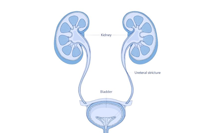
Prevention of ureteral strictures is not always possible, especially in congenital cases, but early treatment of kidney stones, prompt management of infections, and careful monitoring after surgery or radiation therapy may help reduce risk. Prompt attention to urinary retention will also prevent further damage. Narrowing of the ureter can cause renal damage. A severe ureteral stricture can lead to the inability to urinate, which is a medical emergency, as it can lead to hydronephrosis (kidney swelling) and cause the kidneys to stop working (kidney failure).

Georgia Urology is a “Top Doc” independent urology practice, offering exceptional care for men, women, and children. With over 80 providers across more than 30 clinics and ambulatory surgery centers in the Atlanta area, and a 24-hour emergency kidney stone hotline, our goal is to deliver convenient and accessible care to our patients.

FAQs

Will I need surgery?

Not always, as some people may have no symptoms initially. If the stricture is asymptomatic, does not affect kidney function, or is caused by a tumor that responds to other treatments, doctors may opt for observation. Some strictures can be managed with stents, but more severe or persistent cases may require surgical repair.

How serious is a ureteral stricture?

If left untreated, it can lead to kidney damage and become fatal. With proper treatment, outcomes are generally very good.

How long does a ureteral stent stay in place?

Stents may remain in place for weeks to months, depending on the situation, and sometimes require periodic replacement.

Can ureteral strictures come back?

In some cases, strictures can recur, which is why follow-up care is important. Scar tissue that reforms is typically a primary cause.

Ronald Anglade, MD

This page was reviewed by Ronald Anglade, MD

Dr. Anglade is a native of New York. He received his undergraduate degree from Brown University, Providence, RI. He then returned to New York and received his medical degree from the State University of New York Health Science Center at Brooklyn (Downstate Medical Center).

Schedule an Appointment