Pediatric Urology Glossary
A
Ambiguous genitalia – a condition in which a genetic male may have genitals with female characteristics, or a genetic female may have genitals with male characteristics.
Analgesic – any drug intended to alleviate pain.
B
Biopsy – a procedure in which tissue samples are removed (with a needle or during surgery) from the body for examination under a microscope; to determine if cancer or other abnormal cells are present.
Bladder – a triangle-shaped, hollow organ located in the lower abdomen that holds urine.
Bladder augmentation – A surgical procedure in which intestine or other organs are used to make the bladder bigger, usually in cases of spina bifida and severe neurogenic bladder.
C
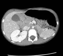
CAT scan: Also called a CT scan. This stands for computerized axial tomography. This enables urologists and radiologists to see cross-sectional images (x-rays) of a patient’s body. See image below:
Circumcision – a surgical procedure to remove the skin covering the end of the penis, called the foreskin.
Cystitis: a bladder infection.
Cystoscopy: The use of a small scope to look inside the urethra and bladder for diagnostic and therapeutic purposes.
Chordee: This is an abnormal bend to the penis which may cause difficulties with intercourse or urination. This anomaly is sometimes only apparent during erections. Boys with chordee also sometimes have hypospadias.
Cryptorchidism: At least one testicle absent from scrotum. The missing” testicle may not exist, or it could be located elsewhere, such as the abdomen. See orchidopexy and undescended testicle.
D
DSD: Disorders or sexual differentiation. See also ambiguous genitalia.
DMSA: This nuclear imaging study determines kidney function and scarring. Radiation is extremely minimal and is extremely safe. See image below:

Diverticulum: outpouching of the bladder, usually due to an area of muscular weakness within the bladder, maybe inconsequential or may require surgery.
E
Encopresis: involuntary and uncontrollable bowel movements
Enuresis: bedwetting
Epididymo-orchitis: infection or inflammation of the testicle and surrounding structures. This is painful, requires antibiotics, and may take days to weeks to improve.
Epispadias: This is the condition in which the urine hole (meatus) is located on the top of the penis, rather than the tip.
ESWL: This stands for extra-corporeal shock wave lithotripsy. This is a method of fragmenting stones in the urinary tract by invisible shock waves. There is no incision and no scopes are used.
Exstrophy of the bladder – the bladder is essentially inside out and exposed on the outside of the abdomen. Because the bladder and other structures are exposed to the outside of the body, urine constantly trickles onto the skin causing local irritation.
H
Hematuria: the presence of blood in urine. This may be microscopic or gross (i.e. urine is pink or red).
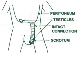
Hernia: a communication between the abdomen and scrotum that enables intestine or other abdominal structures to track into the scrotum. See image below:
Horseshoe kidney – as the kidneys of the fetus arise from the pelvic area they abnormally fuse together at the lower end or base. By fusing, they form a “U” shape, which gives it the name “horseshoe.”
Hydrocele: Fluid around the testicle(s) in the scrotum. This may be due to a communication between the abdominal cavity and the scrotum which may cause fluid to accumulate in the scrotum. See image below:

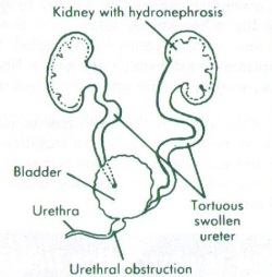
Hydronephrosis: Condition in which one or both kidneys is/are dilated (swollen) with urine. This may be an inconsequential finding on a radiographic test, or it may be indicative of obstruction. Many different pathologic and physiologic processes may be associated with hydronephrosis. See image below:
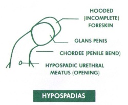
Hypospadias: This is the condition in which the urine hole (meatus) is located on the underside of the penis. It may be located toward the tip (distal) or toward the scrotum (proximal). See image below:
I
Inguinal hernia – when a section of intestine protrudes through a weakness in the abdominal muscles in the groin area.
Intravenous pyelogram (IVP ) – a series of x-rays of the kidney, ureters, and bladder with the injection of a contrast dye into the vein – to detect tumors, abnormalities, kidney stones, or any obstructions, and to assess renal blood flow.
K
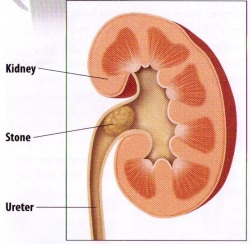
Kidney stone – a solid piece of material that forms from crystallization of excreted substances in the urine.
Kidney transplantation – a procedure that places a healthy kidney from one person into a recipient’s body.
Kidneys – a pair of bean-shaped organs located below the ribs toward the middle of the back.
L
Laparoscopy: A surgical technique which utilizes scopes and cameras to minimize incision length.
Lithotripsy: A method of breaking up a stone in the urinary tract. Frequently used interchangeably with ESWL (see ESWL).
M
MAG-3: this nuclear imaging study determines kidney function as well as the presence or absence of an obstruction. Radiation is extremely minimal and is extremely safe.
Meatal Stenosis: narrowing of the meatus, which may cause pain or difficulty in directing the urine stream.
Meatotomy: a minor procedure performed to correct meatal stenosis.
Meatus: urethral opening or pee hole.”
megaureter – an expanded or widened ureter that does not function normally. The size of a megaureter is usually greater than 7 millimeters in diameter.
Micropenis – a normally structured penis that is below the normal size range for an infant.
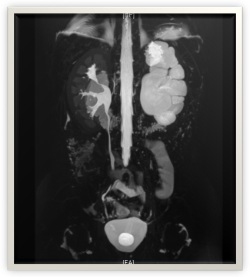
MRI: Magnetic Resonance Imaging. An imaging technique that entails no radiation exposure. However, sedation may be necessary for young children. See image below:
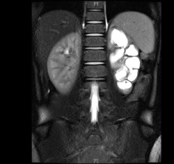
MRU: Magnetic Resonance Urogram. See MRI; Specific type of MRI which gives urologist anatomic detail about the urinary tract. Sedation may be necessary for young children, but there is no radiation exposure. See image below:
Multicystic dysplastic kidney (MCDK): Condition in which entire kidney consists of cysts. These kidneys have no function and should shrink to a minimal size early in childhood.
N
Neurogenic bladder: Condition in which the nerves leading to, or within the urinary bladder cause the organ to malfunction.
Nephrectomy: Surgical removal of a kidney.
Nephrostomy tube: Also called percutaneous nephrostomy tube. Small tube placed into the kidney through the back in order to drain urine.
O
Orchidopexy: surgical procedure that relocates testicle from groin or abdomen into the scrotum.
Orchiectomy: Surgical removal of a testicle
P
Paraphimosis: This occurs after the foreskin has been retracted, but cannot be pulled back over the head of the penis (glans penis) to cover this structure. This is a painful condition and requires immediate attention.
Phimosis: This is when the penile foreskin cannot be retracted (pulled back) adequately in a boy. Some degree of phimosis may be normal, especially in young boys.
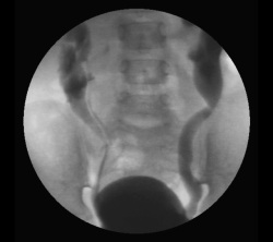
Posterior urethral valves: a condition in which leaflets of urethral tissue cause a blockage in the urethra. This condition requires surgical correction early in life. See image below:

Priapism: Sustained and usually painful erection lasting more than 6 hours. In children, sickle cell disease is the most common cause of priapism.
proteinuria – large amounts of protein in the urine.
Prune belly syndrome – a triad of symptoms that include multiple urinary tract abnormalities. The common abnormalities include an absence of abdominal muscles, undescended testes, and abnormalities of the upper urinary tract.
Pyelonephritis: infection of the kidney, usually including fevers.
Pyeloplasty: a surgical procedure used to correct blockage between kidney and ureter.
R
Reflux: Also called vesicoureteral reflux or VUR. This is when urine backs up from the bladder into one or both ureters. This is very different from acid reflux” or GERD, which involve the flow of stomach contents into the esophagus.
Retrograde Pyelogram (RPG”): X-ray test performed by a urologist that gives valuable anatomic and functional detail about the ureter(s) and kidney(s).
Robotic surgery: Technique in which surgeon controls robotic instruments to perform the surgical procedure.
S
Skin bridge: This is the condition in which a bridge of skin forms between the penile shaft and the penile head after a circumcision.
Stent: There are many kinds of stents used by many different medical and surgical specialists. The stents that urologists place are used to aid in urinary drainage from the kidney to the bladder. These stents are always temporary, and must never be left in place for more than a few months.
T
Testicular torsion: (“torsion”) This is the condition in which the blood supply to the testicle twists, usually resulting in acute and severe pain. This condition requires immediate attention, or testicular loss is virtually certain.
U
UDS (urodynamic study): This test is performed by a urologist to determine causes of abnormal voiding patterns, and to determine whether a patient’s kidneys are at risk of damage from voiding dysfunction.
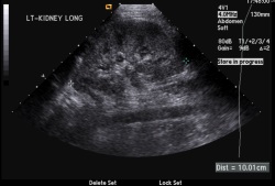
Ultrasound: an imaging technique for visualizing kidney, bladder, testicles, and other anatomies. No radiation. Completely safe. See image below:
Undescended testicle: A testicle that has not descended into the scrotum, as is supposed to occur by 6 months of age. May require orchidopexy.
UPJ obstruction (Ureteral Pelvic Junction obstruction): A congenital condition in which a blockage exists between the kidney and the ureter. May require pyeloplasty. See image below:
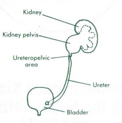
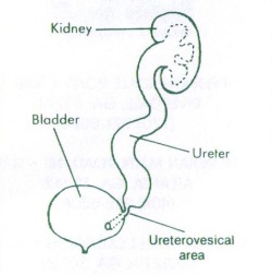
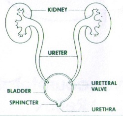
Ureter: A tube that drains urine from the kidney to the bladder. Each kidney has its own ureter, so most individuals have a left and a right ureter. See image below:

Ureterocele: abnormal connection of the ureter into the bladder which may cause obstruction and require surgical correction.
Ureteral reimplant: a surgical procedure used to treat UVJO or urinary reflux.
Ureteroscopy: a procedure in which a small scope is advanced through the urethra and into the ureter in order to diagnose or treat a disorder, especially a ureteral or kidney stone.
Urolithiasis: Condition in which stones (calculi) exist in the urinary tract. They may or may not cause obstruction or pain, and may be located in the kidney, ureter, bladder, or urethra. See image below:
Urologist: Physician specializing in surgical and non-surgical diseases of the male and female urinary and reproductive tracts.
UVJO: Ureteral Vesical Junction Obstruction. Congenital condition in which a blockage exists between the ureter and bladder. This may require a ureteral reimplant. See image below:
Urethra: a tube through which the bladder drains urine. In the male, the urethra is contained within the penis. In the female, this urethral opening is just in front of the vaginal opening and may be difficult for a non-urologist to visualize. See image below:
V
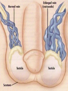
Varicocele: a dilated group of veins draining the testicle, usually located on the left side. This can be associated with infertility, testicular growth retardation, and rarely a dull pain. See image below:
VCUG: This is an abbreviation for voiding cystourethrogram. This is a test performed by a radiologist which tests for urinary reflux and other bladder anomalies. It is an x-ray that requires that a urethral catheter be placed temporarily. See image below:
Vesicoureteral reflux: see reflux.
Void: to urinate
Voiding dysfunction: abnormal or pathologic storage or elimination of urine.